Michelle L. Ward BSc BVSc(Hons I) CertZooMed MRCVS
Exotic Animal and Wildlife Service, Royal (Dick) School of Veterinary Studies, University of Edinburgh’s Hospital for Small Animals, Easter Bush Veterinary Centre, Roslin, Midlothian, EH25 9RG
Based on a presentation to the Northern Symposium at Chester Zoo on 21st October 2006
Chelonia comprise some of the most fascinating creatures on the planet. Growing up in suburban Sydney in Australia I have distinct childhood memories of hours spent watching the captivating activities of our local wild chelonian, Chelodina longicollis. More commonly known as the long-necked tortoise or the Eastern snake-necked turtle, this freshwater species is a common inhabitant of creeks and other waterways in the region. Watching them forage for aquatic invertebrates, tadpoles and small ish whilst sitting on a rock above the clear waters was a favourite pastime. Despite my interest, I was always pleased to keep a healthy distance to avoid being exposed to the characteristically malodorous secretions that are produced by glands in their axillae (‘armpits’) and groin. Their agility in the water, their relaxed attitude whilst basking on land and their unpleasant defence mechanism were all part of the attraction.
It has become clear to me over the years that I am not alone in my fascination with chelonia. On arriving in Britain in 2001 to work in general veterinary practice I was astonished at the number of pet tortoises that were planted on my consultation table each week. Working with Mediterranean tortoises, in particular, was a new and wonderful experience for me and it became apparent that the British public had fallen for these creatures too. More recently, at least in the areas of the country where I have worked, pet terrapins and turtles appear to be increasing in popularity also.
As with all animals, the keeping of chelonia as pets carries a number of responsibilities. One of those is to protect the animal from ill health. Ensuring optimal environmental and dietary conditions as well as protecting the animal from trauma and predation are key factors in the maintenance of good health. The most common diseases seen in pet chelonia at the present time occur due to inappropriate husbandry conditions, often inadvertently provided by their owners. Veterinary surgeons have an important role to play in assessing health requirements, detecting potential problems and advising on general care, husbandry alterations and/or treatment.
Veterinary care
Traditionally chelonia were taken to the vet only when they became injured or ill. In addition to providing vital treatment at times of known disease, it is now accepted that all exotic pet species beneit from routine health examinations as well.
Challenges for the veterinary surgeon
As an exotic animal vet, the opportunity to work with chelonia on a regular basis is a thrill. However, there are a number of challenges that need to be overcome to permit a thorough assessment and safe treatment.
First and foremost, the evolutionary adaptation of the shell not only protects the animal from predators; it also protects it from the prodding ingers of the vet! The shell obviously restricts access to the internal organs and this can have some implications for clinical assessment. Sometimes this obstacle can be overcome easily. For example, it is almost impossible to use a standard stethoscope to count the heart rate of a chelonian. Instead the use of a Doppler probe allows measurement of heart or pulse rate; vitally important parameters for assessment of a collapsed or anaesthetised patient.
The second challenge of note is the range of species that a vet may be presented with. A wide variety of species may be kept as pets and each has its own speciic needs and disease susceptibilities. For the rarer species sometimes very little is known about their requirements. Despite a broadly similar external appearance, terrestrial and aquatic species are vastly different in their husbandry requirements and this may have implications for how disease can be treated. And, of course, the size of the patient can vary enormously (Fig. 1). It is not uncommon in our hospital to have a newly purchased pair of red-eared terrapin (Trachemys scripta elegans) hatchlings weighing less than 5g each, a 1.5kg Hermann’s tortoise (Testudo hermanni) and a 20kg African spurred tortoise (Geochelone sulcata) in for assessment all on the same day.
Metabolic rate is variable from species to species with larger individuals and herbivores having a slower metabolism than smaller individuals and omnivorous or carnivorous species, respectively. In general it is true to say that reptiles have a slower metabolism than mammals and that their metabolism is dependent on the ambient temperature. This is important because the drugs that may be prescribed are generally human or domestic mammal medications. The dose often has to be altered accordingly and the temperature of the environment taken into account when calculating dose frequency.
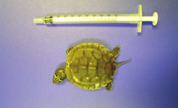
Fig. 1. Chelonian patients can be very small indeed. This terrapin hatchling was presented for assessment of the focal white patches on the carapace. A bacterial infection was diagnosed by taking samples of the lesions and assessing them under a microscope. The problem was treated successfully with antibiotic ointment and by improving the water quality in the tank.(courtesy of Michelle Ward.)
A particular challenge when using anaesthesia for diagnostic or surgical procedures is that some chelonian species have the ability to hold their breath for prolonged periods. Mechanical ventilation can overcome this problem.
When dealing with wounds on aquatic species, treatment or husbandry may need to be modiied to account for the animal’s normal lifestyle. Many wounds heal better if exposed to the air so it may be necessary to restrict access to the water for limited periods. In addition, bandage materials do not provide suficient protection if they are constantly immersed in water so innovative methods for keeping wounds clean need to be devised.
Finally, if chelonia develop a localised infection or abscess anywhere on their body, their pus, unlike that of most mammals, forms a solid plug. This is similar to the situation in birds and the treatment of choice is usually surgical removal. Lancing and draining an abscess, as is performed frequently in cats and farm animals, does not resolve chelonian abscesses.
Injuries
Common injuries that occur in pet chelonia include dog or fox bites (Fig. 2), rat bites (during hibernation), burns (due to inappropriate heat sources), car or lawnmower injuries, fractures (e.g. due to metabolic bone disease), damage to the nails from inappropriate substrates or constant attempts to escape, aggression from cage or tank mates and trauma during mating.
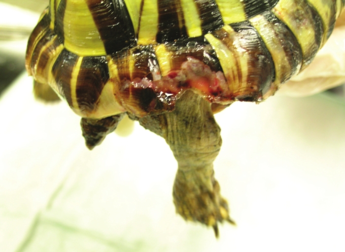
Fig. 2. A shell injury caused by a fox bite in this young Testudo hermanni. The injury was lushed and cleaned. Bandages were applied to protect the damaged tissue.(courtesy of Michelle Ward.)
Illness
Broadly speaking, there are two types of illness that may be observed in pet chelonia. The irst is when the symptoms or clinical signs are quite obvious and direct the vet to the cause of the problem. Examples include lameness, hemipenile prolapse, convulsions, oedema (swelling due to luid accumulation in one or more parts of the body) or regurgitation.
The second and much more common scenario is the tortoise, terrapin or turtle with very vague symptoms. Reduced appetite, anorexia, lethargy and/or altered behaviour are some examples. Almost all diseases can cause these signs. Often the signs seem very subtle when, in fact, the animal may be seriously unwell. Like their wild counterparts, pet chelonia are skilled at masking signs of disease, a feature which minimises the risk of predation. So, even in a captive situation, problems are often detected only once the disease is in quite an advanced state and the animal no longer has the ability to hide it. This can make accurate diagnosis and treatment much more challenging.
Examples of common diseases
Shell, skin and eye problems are relatively common. This is probably because they are easier to detect than disease of internal organs.
Shell rot refers to infection of the shell with bacteria, fungi or algae. If caught early, treatment is usually successful and involves the removal of the infected tissue and topical medication. Sometimes systemic medication may be prescribed.
Sloughing of the skin can occur due to local skin infections, poor nutrition (e.g. low vitamin A levels), access to water with a high bacterial load and/or inadequate haul out areas for aquatic species. A severe case that resulted from generalised septicaemia is shown in Figure 3.
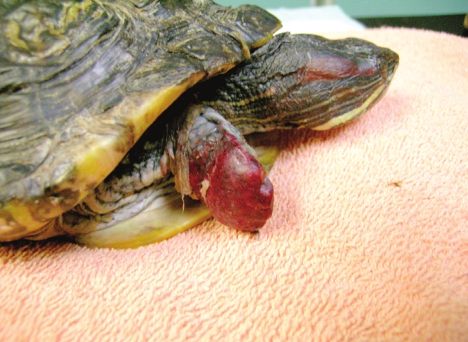
Fig. 3. Complete sloughing of the skin from the right forelimb of this Trachemys scripta elegans suffering from septicaemia. This animal was euthanased due to the severe nature of the disease.(courtesy of Michelle Ward.)
Diseases of the eye may occur due to infection, trauma, fat deposits, frost damage during hibernation, the presence of foreign material and nutritional deiciencies. Assessment and treatment of the eye is similar to that in domestic species.
Reproductive disease is also relatively common. In females this is most often in the form of dystocia (retained eggs). Dystocia may occur as a result of adverse environmental factors, mechanical obstruction (e.g. pelvic damage or an abnormally large or malformed egg), generalised illness or a hormone problem. The vast majority of cases can be resolved by providing a suitable nesting area and an appropriate environment. If this is unsuccessful, medical therapy can be attempted. Finally, surgery may be considered for cases that cannot be resolved by other methods. In males prolapse of the penis may occur due to generalised weakness, nerve damage, infection, mating injury or secondary to straining e.g. due to a bladder stone or an intestinal obstruction.
Routine health checks
Routine veterinary health checks are highly recommended for all pet chelonia. Dogs, cats, rabbits and ferrets visit the vet for check-ups on a regular basis for vaccination. A full health check is usually performed at the same time. Although we do not vaccinate tortoises, terrapins or turtles, to ensure optimal health, they too should be seen by a vet on a regular basis for health assessment. For most pets this includes an initial appointment when irst acquired and then regular reviews e.g. once a year. In addition to a full physical examination, these appointments provide an opportunity to discuss the pet’s behaviour, husbandry, general health and whether any disease screening is warranted. For clients with new animals, advice can be given regarding appropriate quarantine to protect other reptiles within the household from infectious diseases.
For terrestrial species that originate from temperate climates, the ideal time for an annual health check is prior to hibernation. This is because hibernation can exacerbate underlying medical problems. Many post-hibernation problems are due to illness that was present prior to hibernation. At our hospital we host a pre-hibernation evening each October. All the clients with tortoises registered with the practice are invited to attend. Presentations are given to update attendees on the latest discoveries in chelonian medicine and to review hibernation recommendations. Each tortoise is given a free health check and, when possible, the females have a reproductive tract ultrasound scan to assess the activity levels of their ovaries (Fig. 4). These evenings are a fun way for owners to meet other tortoise enthusiasts, to ask questions about health and hibernation issues and to ensure their pet is healthy prior to hibernation. Regardless of whether a particular practice organises such events, a full health check with a local vet at this time is worthwhile and provides peace of mind over the winter. Equally for species that do not hibernate, an annual health exam is valuable but the timing of such an appointment is not so important.
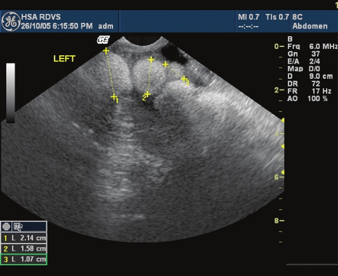
Fig. 4. An ultrasound scan image from a female Testudo graeca performed during a pre-hibernation health check. The follicles on the ovaries are seen as white circular structures at the top of the image. The presence of follicles of varying size suggests normal ovarian activity in this animal.(courtesy of Regine Hagen.)
Diagnostic tools
A full physical examination is very useful and provides a vet with much information regarding the animal’s health. However, it has been previously mentioned that chelonia can masterfully hide signs of disease and that their unique anatomy places some limitations on a health assessment based purely on a physical examination. Therefore there are situations when one or more diagnostic tests may be suggested as a means for more thorough health evaluation. The diagnosis of disease of chelonia is becoming increasingly sophisticated and this is having a positive effect on the health of the pet population. Some of these tools will be discussed briely below.
Faecal tests are useful to detect parasite burdens or microbial imbalance within the gut. Samples are usually easy to collect and the tests are generally inexpensive.
A blood sample can be safely collected from all but the smallest of chelonia. Even 0.5ml of blood can provide an immense amount of information about an animal’s health. For general health screens two types of blood tests are carried out.
Haematology (also known as a complete blood count or CBC) assesses the number of and characteristics of the red blood cells and the various types of white blood cells in the animal’s circulation. This may provide clues regarding immune status, presence/absence of infection, presence/absence of blood parasites, stress levels and degree of dehydration or blood loss. Leukaemia is an uncommon disease in chelonia but can be detected on haematology.
Biochemistry assesses the levels of various other compounds (e.g. enzymes) in the circulation. This may provide information regarding the health of the kidneys, liver and muscle as well as assessing possible nutritional deiciencies.
Various samples may be assessed microscopically. Discharges from the eyes or nose or aspirates from lumps on the body may be evaluated under a microscope to determine the most likely cause and therefore the best course of treatment. In this way it may be possible to detect bacterial infection, tumour cells or fatty tissues.
Virology describes the testing of samples for the presence of a speciic virus. The most common chelonian virus screening test requested at our hospital is the PCR test for Chelonian herpes virus. This particular virus can cause chronic health problems. A nasal lush or a sample of nasal discharge is examined in a laboratory for the presence of the virus particles.
Diagnostic imaging is incredibly useful for evaluating the health of both small and large captive chelonia. This includes tests such as radiography (xrays), ultrasound, endoscopy, CT (computed tomography) scans and MRI (magnetic resonance imaging) scans.
Radiography is the most common diagnostic imaging modality utilised in veterinary practice. Most clinics will have an xray unit that is used for domestic animals that can be successfully employed for imaging tortoises, terrapins and turtles. Xrays may be taken of the whole body (usually three different views) or just of one area of interest e.g. a suspected fracture. Radiography is particularly useful for assessing the health of the bony structures of the body (e.g. for diagnosing metabolic bone disease, dislocations or bone infections) and the lung ields (e.g. for diagnosing pneumonia). Additionally, ingested stones or metallic objects (Fig. 5) and shelled eggs within the reproductive tract are easy to detect on xray.
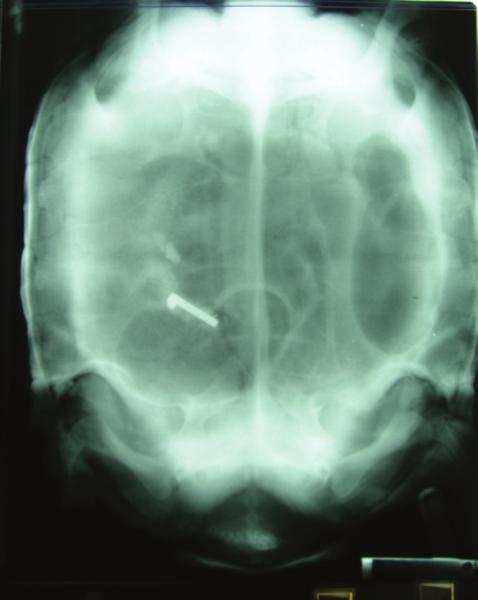
Fig. 5. This dorsoventral xray of a chronically anorexic Geochelone sulcata reveals a white metallic object in the intestines. There is also a large pocket of gas (darker kidney-shaped area) around the foreign object and another in the stomach (on the right of the image).(courtesy of Michelle Ward.)
Ultrasonography is useful for evaluating the internal organs of the coelomic cavity including the heart, liver, ovaries, stomach and intestines. A machine similar to that used for pregnancy scans in people is used. It is not possible to scan through the shell so the probe is placed in front of one of the legs and angled toward the inside of the body. Examples of health problems that can be detected on ultrasonography include pre-ovulatory follicular stasis, ovarian cysts, liver disease, internal masses (e.g. abscesses or tumours) and coelomitis (the reptilian equivalent of peritonitis). The procedure is not painful and can be performed easily in most chelonia. Sedation may be required in very large, powerful animals or in those with a hinge. Ultrasonography can also be used to assess disease of the eye or to evaluate the extent of a mass on the skin.
Endoscopy describes the use of small cameras that are itted within a rod-like instrument to visualise the internal organs of an animal. Both rigid and lexible endoscopes are used for different purposes and there are various sizes available. The equipment is expensive and delicate and is not available at all veterinary practices. Endoscopes can be used to look inside the coelomic cavity (entering in the pre-femoral fossa, i.e. in front of the back leg) to view the internal organs directly or can be introduced through the mouth to enter the respiratory or upper gastrointestinal tract (Fig. 6). Similarly, the endoscope can enter the body via the cloaca to visualise the bladder, colon or reproductive tract. The technology of endoscopy is advancing all the time and improved models are being developed to improve the clarity of the images obtained and the function of the equipment. It is now possible to not only look at or within the organs of interest but also to take samples (e.g. biopsies) or to remove foreign objects via portals in the equipment. Sedation or anaesthesia is generally required for endoscopy.
Computed tomography. (CT) and magnetic. resonance. imaging. (MRI) are advanced imaging tools that are uncommonly used in chelonian veterinary practice at the present time. Very few practices have access to the equipment and the cost is prohibitive for most owners. However, the three-dimensional evaluation of tissues that can be achieved allows for incredible detail to be gained about patient’s organs in a non-invasive fashion. It is hoped that in the future these tools will become more widely available and less expensive to permit evaluation of chelonian and other veterinary patients.
Finally, surgical exploration may be considered as a diagnostic tool in complicated or dificult cases. This is uncommon but can be helpful if an ill animal is failing to respond to standard treatment. If a problem is identiied as the source of illness during surgical exploration, often treatment can be instituted at the same time, e.g. removal of a foreign body or mass.
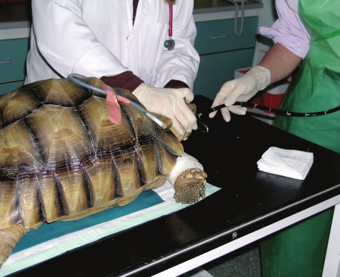
Fig. 6. A lexible endoscope is being introduced into the stomach of this Geochelone sulcata who had been regurgitating blood for a few days. A stomach ulcer was diagnosed via endoscopy and the tortoise made a full recovery following a course of medication used to treat similar ulcers in humans.(courtesy of Michelle Ward.)
Summary
Pet tortoises, terrapins and turtles are popular pets that deserve first class veterinary care. Our understanding of their needs and disease susceptibilities is improving all the time. It is important that these advances in knowledge are disseminated to the chelonian owning public so that their pets can reap the beneits.
Routine examinations, usually arranged on an annual basis, are highly recommended for evaluation of general health status and to discuss husbandry and behaviour issues. A range of diagnostic tools is available that can assist in the investigation of disease if health issues arise. Early diagnosis is often the key to successful treatment.
It is hoped that with improved understanding of environmental needs and more sophisticated health monitoring tools, the incidence of husbandry-related illness will decrease in the future. The keeping of chelonia as pets is a privilege that brings enormous pleasure to many people. In return, it is our responsibility to ensure the health of the animals that ind themselves in our care is maintained at the highest level.
Testudo Volume Six Number Four 2007
Top